Career Update: Part 1

So I started out in Cardiac Telemetry as a new grad. I knew right out of school that I wanted to have  Telemetry as a foundation for my practice. If you are starting out as a new grad in the US, Tele is a great start. From Tele it is much easier to navigate into specialty areas than moving from Med-Surg. After about 18 months I was fatigued with my job. I worked on the biggest unit in the hospital which was split into regular Tele, Med-Tele, VIP unit, and observation unit; as well as being floated to MICU, CVSD, Maternity, Med-surg and the new sister hospital which had opened a good 20 min drive from the main campus. I had started working on days as a new grad as I thrive under pressure. I learned so much in the first 6 months and definitely found my groove, understanding my patients, documentation and the docs. I found that I really enjoyed working the observation unit as it was high turn over, fast paced, and a bit of a mystery of what the ER was bringing up. One time I had a pt come up dx: Chest Pain. I gave her morphine, did an EKG, put her on O2, call the doc – she was immediately sent to the cath lab. This pt should have been a cardiac alert, turned out that she had 100% occlusion in the Circumflex – she was dying. I loved that I was on the cusp of something, it was fast, it was more challenging than the other units and I liked it. After about 14 months on days, I switched to night shift. Our night shift staff was thin, a lot had left and they were struggling, and to be honest, I needed the pay increase. So I did nights for the last 5 months of my job and started looking for other opportunities.

I landed a job at another hospital, again it was a non-profit hospital, but in an affluent area. I was working in CV-Step Down, sister floor to CVICU post-CABG/TAVR pts mainly. I was thrilled that I was moving deeper into the cardiac specialty, this is what I wanted. After 3 months in my new job I wasn’t happy. I was working nights, the staff were burned out and it showed. I found that the acuity of my patients weren’t all that different from my prior job. Post-open heart really only meant, possible Afib – start Cardizem or Amiodarone drip, chest tubes – usually 2-4, Dermabond midline chest incision and leg donor site, and lastly a foley. There was nothing really interesting about it. The fun happened in the OR, ICU and then during the day when the chest tubes were pulled, I missed out on all of that and just ended up changing the chest tube dressing and drawing blood from the central line.

I started to feel depressed; I started dreading going to work. I wondered if I was looking for something better that didn’t exist. I thought nursing was this great expanse of possibilities, but here I was feeling crappy after changing jobs from a crappy position to another one. I considered if I just wasn’t made for nursing. I’ll be honest, I never planned on being a nurse, I didn’t grow up dressing up as a nurse and putting a stethoscope to my teddy bear – that wasn’t me. Life brought me to nursing, and my ability to take on a challenge, think critically, enjoy interacting with people, had made this a good choice for me, but now I was here in this place where I was unhappy and couldn’t quite figure out how to fix it. So I did what I thought I should do and start looking for another job. Now I was looking for a higher level: ER, ICU, ICU specialities.

Ironically I reapplied for some of the jobs I had applied for when leaving my first job. I really just felt like I had nothing to lose. I did get a call back for an ER job at a Level II Trauma hospital right on the highway. It is one hospital within a large hospital system, which means there are a lot of jobs available without actually quitting and having to learn a whole new system over again. So, I booked the interview and figured I’d wing it, at the end of the day, I still had a job, even if I didn’t get a new one, the bills would still get paid haha!

Stay Tuned for what happens next…!


If you like this post, please press like below! 🙂

EKG: Hypokalemia vs Hyperkalemia

So you should already know that potassium (K+) is VERY important in our bodies. From the action of K+ in the depolarization and repolarization which takes place in the heart, you can imagine that if there is too little, or too much potassium, then the effects will be present in the heart muscle.

HYPOkalemia

Hypo –  meaning; less, not enough, below normal levels. Therefore, here we are talking about not enough potassium. Below is an EKG, and from my last posting where you became familiar with a normal EKG, you should be able to notice there are things about this EKG that are abnormal (If you noticed that, then well done!)

You can see that the U wave is really big, or in medical terms; prominent U waves are present. The T wave on the other hand is said to be “inverted” or the opposite direction that it should be, it can be flattened also. So just looking at this portion we can see that the REpolarization phase is definitely going to be affected by these changes.

I like to think of hypokalemia as making everything more depressed. A depressed person, moves slower, things take longer for them to do, they are disorganized and one thing runs into another.

Other things that can be seen in Hypokalemia in the EKG: the ST segment can be depressed (below the flat baseline), the PR interval can be prolonged (longer that should be) as well as the QT interval (due to the merging of T and U waves) and the P wave can get taller and longer.

Hypokalemia is serious because it can develop into life threatening arrhythmia such as Torsades de Pointe, Ventricular Tachycardia, Ventricular Fibrillation.

HYPERkalemia

This is the opposite, HYPER: too much, more than baseline, overload. Too much is just as dangerous and not enough, and as you can see from the strip below, the EKG will show you signs of high K+.

P waves here in this strip is missing; you might see small to no P waves, coupled with spiked T waves. Look at the T waves here, they are as tall as the QRS complex! In severe hyperkalemia, the QRS complex can widen also.

So as with hypokalemia, I like to think use little story concepts to remember the differences. So I like to think of hyperkalemia as an ADHD child: the T waves are hyper and the P waves are playing hide-&-go-seek. Repolarization period here is affected by surplus potassium by causing a faster repolarization period, and reducing the response of sodium channels in the myocardium; therefore slowing conduction around the heart and reducing the P wave in the strip.

So how can we tell when someone is hyperkalemic? Think: Blood test (straight forward, check K+ level), ABG (metabolic acidosis), hx renal disease (reduces elimination of K+), Addison’s disease, severe burns (mass K+ being released for cells/cell-lysis?), Digoxin toxicity.

What to know about potassium:

Potassium is mostly found inside the cells.

The normal range for potassium is 3.5-5.0 mEq/L

Cardiac Tamponade

This has nothing to do with tampons in the heart ok! Just an FYI.

When blood or fluid accumulate in the sac which surrounds the heart it exerts pressure against the heart, making it harder for the heart to pump. As you can imagine, if the ventricles cannot fill fully or contract effectively then cardiac output decreases. Decrease cardiac output is not good news!

So how do you know if someone has this?

Well the symptoms are nothing unusual: chest pain, restlessness, quick shallow breathing, palpitations, pallor, tachycardia, weak peripheral pulses, pulsus paradoxical, distended neck veins, low BP… (do these all sound a little familiar? MI anyone?) Well causes of cardiac tamponade can be MI, Dissecting aortic aneurysm (thoracic), end-stage lung cancer, heart surgery, Pericarditis caused by bacterial or viral infections, or direct wounds to the heart.

How do we diagnose? Echocardiogram is the usual diagnostic tool, but MRI, CT, CXR or EKG can also highlight or confirm this.

cardiac-tamponade-treatment-s48jtsnc

Can you see the halo of fluid around the heart?

This is an emergency! The fluid must be drained to reduce damage to the myocardium and reestablish cardiac output.

Pericardiocentesis is a procedure that uses a needle to remove fluid from the pericardial sac, the tissue that surrounds the heart. The patient will be hyperoxygenated, given fluids to maintain blood pressure. It is important to note that a pericardiocentesis may have to be repeated because cardiac tamponade can return. With fast treatment the outcome is usual good, without treatment death is immanent.

A pericardial draining tube may be placed during surgery to keep in place postop. The drainage will be measured every shift and documentation on the color, amount and consistency is very important! The tube will usually be removed slowly over the postop recovery weeks at the discretion of the cardiologist.

Nursing Skills: Reasons for Chest Tubes

So I thought I’d write about chest tubes this week because I had a patient with two chest tubes and was post removal of a pericardial tube also. Not only that, but a colleague had a young pt in his 20’s whose chest tube popped out on the same day! Needless to say I thought it would be a great topic to cover. I’ll break it into three postings

Firstly, chest tubes aren’t as scary as they appear: sure, they are stuck in someones chest but there are many different reasons for a chest tube so knowing a thorough background on your pt is very important.

The procedure for placing chest tubes is called a Thoracostomy.

Reasons for inserting a chest tube:

Pneumothorax: This is when air collects in the pleural space; can also be referred to as a “collapsed lung”. The air pressure in the pleural space does not allow the lung to reinflate.

Pneumothoraxes can occur after central line insertion, after chest surgery, after trauma to the chest, or after a traumatic airway intubation. It is very important to remember that if the air continues to collect in the chest, the pressure in that pocket can increase and push the whole mediastinum over to the other side – this is called a “tension pneumothorax”, and is  life-threatening.


hemothorax1 Hemothorax: This is is when blood fills/pools in the pleural space and reduces the area for lung expansion. This can happen due to trauma or surgery. There is a possibility of having a hemo-pneumothorax which means air and blood fill the cavity. Also there remains the risk for medialstinal shift “hemo-tension-thorax”, this would be an emergency.


Pleural Effusion: This is the accumulation of fluid in the pleural space. This can be caused by CHF, liver failure, kidney failure, peritoneal dialysis, pneumonia, lymphoma or breast cancer. Though most pleural effusions are caused by congestive heart failure, pneumonia, pulmonary embolism and malignancy.


 Empyema: This is when pus is the substance which fills the pleural space. This is clearly cause by infection, but the pressure of pus in the pleural space makes it difficult for pts to breathe freely, they will likely have fevers and chills, malaise and chest pain.

Hypocalcemia – Got Milk?

These test for hypocalcemia seem so random? Who in the world discovered them? Well, I guess that’s not the most important point here, the fact is: hypocalcemia is very dangerous! Think about the role calcium plays in our muscles and bones? Our heart is a muscle, so you will expect to see changes there if calcium is either low or high. So these signs Chvostek and Trousseau – I guess they are the geniuses who discovered the sx, but seriously? Could they get more complex names?! Ugh! Ok so how I remember the two, is Chvostek is “Cheek”, and Trousseau’s is “Thumb”. I find that this helps me to differentiate between the two.

Major sx to watch for is TETANY – what in the world is that you might be thinking. Well is basically means muscle spasms or twitching. So why does calcium affect muscles in this way? Calcium blocks sodium channels and stops depolarization (contraction) of nerve and muscle fibers, therefore, a lack of calcium reduces the threshold for depolarization, making the muscles jerky.

A mnemonic for hypocalceima sx:

“CATS go numb”- Convulsions, Arrhythmias, Tetany and numbness/parasthesias in hands, feet, around mouth and lips.

So is this just someone who is skipping their milk and cookies at night? No. Hypocalcemia is something seen with an array of diseases. Calcium is found in the bones, joined with other substances or free floating in the blood. Sure people with eating disorders or severe malnutirtion can have this, but it is mostly seen with those with thyroid issues. Parathyroid hormone (PTH) in a healthy person is what tightly controls calcium levels in the body. So how can PTH stop working? Well if I were assessing my patient for hypocalcemia, a crucial question would be any surgery or injury to their neck or head. Why is this? Well damage to the parathyroid in any way can inhibit the function of PTH and hypocalemia ensues. Hypoparathyroidism would be the name for this abnormality in PTH function. Surgical destruction of the parathyroid glands by parathyroidectomy, partial or total thyroidectomy, or neck dissection for head and neck cancers or any autoimmune issue can be a good indicator of why hypocalcemia is presenting.

Great Little Story to Remember Those AV Heart Blocks!

❤The Sad Story of a Struggling Marriage…❤

1st Degree AVB
Husband comes home late every night at the same time, but he
ALWAYS comes home!!!

** P waves aren’t on time but always present! Prolonged PRI

2nd Degree AVB Type 1 (Mobitz 1 or Wenckebach)
Husband comes home later and later and later until one night he
doesn’t come home at all, then the pattern starts all over again!!!

**P wave “Going, going, gone”, PRI gets longer & longer till P is dropped

2nd Degree AVB Type 2 (Mobitz 2)
Husband comes at the same time every night, but there are some
nights that he just doesn’t come home!!!

**P wave is on time but randomly drops

3rd Degree AVB (Complete)
Husband and wife are finally divorced but can’t afford to move out of
their house because of the bad economy. As a result, they are still
living under the same roof, but leading two separate lives!!! It
appears as if they’re still married, but they’re not! There is no
communication between them!!!

** P waves, PRI are not in sync with Ventricular activity (QRS) and its a hot mess!

EKG Time: I never see U waves?

Have you been diligently looking through EKG strips on your Telemetry unit trying to find a u wave? But they taught you in school it was apart of the EKG complex right? Well yes, but they are not seen all the time, which isn’t a bad thing, in fact it doesn’t really matter that much. We don’t talk about U waves very often because there isn’t really much to say, there are usually other “cardinal signs”, where a U wave can confirm something we can already see. Could be classified as last phase of ventricular repolarization or endocardial repolarization, but at the end of the day, we don’t really understand it fully though there are some things you should know about U waves:

  • U waves follow T waves and should be about half the height of the T wave.
  • The U wave should be less than 2 mm (2 tiny boxes on the EKG strip).
  • They are mostly seen or more clearly seen, in sinus bradycardia, but if you can’t see them it’s ok!
  • Best seen in leads V2, V3
  • They can often interfere with the measurement of the QT intervals.

So you’ve found a EKG strip with some really cool U waves, you still need to know what the changes mean. The changes seen in U Waves (either prominent upright or depressed/inverted) are usually correlating to some other clearly visible change in the EKG (T waves changes, PR intervals or ST seg. changes). So what are some changes you should know about?

Inverted U waves are very myocardial focused. They usually mean trouble. Ischemic heart disease (often indicating left main or LAD disease) is one of a few serious readings that can come from a severely inverted U wave; Myocardial infarction (in leads with pathologic Q waves) and during a angina attack (acute ischemia or exercise-induced ischemia) they can also be present.

Upright waves (prominent ones that is) can mean Hypokalemia (remember the triad: ST segment depression, low amplitude T waves, and prominent U waves). There are times in pathological cases where the T and U wave can merge together to create a large wave, it makes it very tricky to know if it is a very large elevated T or a merge, look at the full picture, does this patient have a CNS disease or disease process going on? Then most likely it is a merge. Lastly, drugs. We all know that drugs interfere with all sorts of things, so what drugs could be messing with your U waves? Quinidine and other type 1A antiarrhythmics. Any drug that is acting on the heart can have adverse effects, but don’t forget to think about antipsychotics too! So keep this in mind when reading your EKG. Although you are focusing on a map of the heart, keep in mind that this is a patient, and every patient has a physiological story. The map you are looking at can tell you a snapshot of it’s current mapping system, but there are always other factors to consider.

EKG Time!

Image

EKG TIME! (For basic understanding)

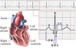
Ok so for many the EKG is a weird and wonderful map of squiggles, but actually each little squiggle is very important in translating the conduction of the heart. From your A&P course you should know that the normal conduction of the heart starts in the SA node in the Right Atrium, it travels to the AV node in the wall of the right atrium (middle of the heart), then through the Bundle of His/Left & Right Bundle Branches and down through the Purkinje fibers. The EKG is basically a map of that scenario. Imagine that you are in a car in San Antonio (SA node), you need to drive to Aventura (AV node), then you go through the spaghetti junction (Bundle of His) while heading to Pittsburg (Purkinje fibers) – if you were to use Google maps, you would have the route highlighted out on the roads you have to take, right? Well that is basically what an EKG is! It is the highlighted route the conduction took through the heart.

I hope that has given you a little clarity on those squiggly lines, haha!

Now what does the map tell us?

As you can see we use letters to signify different areas of the squiggle. You can see that P is a little bump at the beginning. This is showing us that the atria of the heart are depolarizing (another fancy word for compress or pump). The atria is pushing the blood into the ventricles. P shows that the car has left the SA node in the Right Atrium and is traveling to the AV node in the middle of the heart. From P to Q (called PR complex),  the car then leaves the AV node and travels down through the Bundle of His. One the car travels out from the Bundle of His down the Bundle Branches, through the Purkinje fibers and the rest of the heart so a QRS complex occurs. The QRS shows that the ventricles have received all the blood from the atria and pumps it back out to lungs (right V) and aorta (left V). And with that you have just witnessed a heartbeat.

Well what about the leftover squiggles…

So between the S and the T (called the ST segment), we see the heart recovering from the heartbeat, otherwise known as Ventricular REpolarization. Think about clenching your fist really hard and then relaxing, that is in essence what the heart is doing. The ST segment is very important to monitor on an EKG strip because if you are looking for something pathological, such as an MI, you might just find it here with ST segment elevation. This would mean the patient would be diagnosed as having a STEMI. We could continue this further, but we’ll save the pathological changes for another time.

So following the T wave, the heart is now ready to complete another beat! We have come full circle!

I hope you found this helpful, it is purely for basic understanding, EKG’s can be much more in depth! My best advice is so search YouTube for videos, as visualizing this is the best way to learn what it going on inside the heart!

Good Resources:

http://www.youtube.com/watch?v=7b98JcGIGyE

http://handwrittentutorials.com/

Rapid Interpretation of EKG’s 6th Ed. – Dale Durbin (really good book)

Welcome! The Introduction!

Hello One & ALL!

I have created this blog as a way to connect with other RNs and become a resource of learning for nursing students. I remember when I was a nursing student. I knew nothing about nursing. I wasn’t one of those people who was a 5th generation nurse, no-one in my family was medical, or had even finished college to be completely honest. I needed a place to ask questions, I needed to ask “stupid” questions without being judged. Some of the forums I used for a while, but even there I found there were some of those “eat their young” bullies, so I quit using them.

I hope to make this a place where anyone can ask question or learn. If you are thinking about getting into nursing, are a student, or are a well seasoned nurse we all need your questions and your wisdom.

One thing that I have found so beautiful about nursing is the learning element – you really do learn something new every day! It is such a vast and versatile field that there genuinely is something for everyone. In nursing school there were things which I knew flat out was not for me, but I didn’t let it put me off nursing. Nursing school is just one of those things you have to get through, to get to where you want to be. 

So there is my little introduction. I hope this is the beginning of a wonderful journey. Please tell others about it – the more the merrier! I would love to have guest posts in the future from experienced nurses in specialty areas, as well as giveaways etc. I am looking forward to the future of this blog!

Much love,

JaeJ,RN