Nursing Skills: NG Tubes!

NG tubes. Yes, they’re pretty gross. Anything going in or coming out of the nose is gross, have you seen the stuff that comes out when an NGT is put to suction? I’m talking rainbow secretions! All joking aside, if you think they are gross, just imagine how the patient feels. I put a NG tube in the other day and was struggling to get it around the sinuses, it isn’t a straight route y’know.

So advice for NG insertion:

  • Explain EVERYTHING to the patient! Tell them it will be uncomfortable. Tell them you will be there for them. Tell them that it is crucial to swallow. Tell them you are going to try to do the procedure as quick as possible. REASSURANCE is key!
  • Pre-medicate if necessary. This is controversial. My patient the other day got morphine sulfate IV before the procedure, this totally helped in chilling out her gag-reflex and made the process easier BUT, if your patient is prone to becoming sedated or unable to follow commands then lay off the meds – use your judgement.
  • Measure correctly. Remember that it is nose to ear to xyphoid process – mark it with tape.
  • Lube, lube and more lube! We need that tube to slide and it has a long way to go, so be liberal with that lube!
  • Encourage your patient to sip water. The swallowing effect with assist you in moving the tube down the esophagus.
  • Don’t give up in getting it down! There may be issues along the way so troubleshoot fast. If you cannot get it pass the sinuses, twist and manipulate the directing you are inserting the tube. Have the patient open their mouth to check for coiling in the back of the throat.
  • Lastly, always check your placement. Use the piston to blow an air bubble in, listen at the xyphoid process with your stethoscope for the air bubble. Placement is also checked with a follow up CXR.

There are many reasons why an NG tube may be placed. There are reasons to suction, give meds, remove acid, or feeding – regardless they are not comfortable for the patient and careful consideration should be made for skin breakdown in the nares or insertion site.
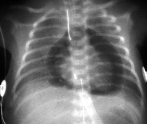
Cardiac Tamponade

This has nothing to do with tampons in the heart ok! Just an FYI.

When blood or fluid accumulate in the sac which surrounds the heart it exerts pressure against the heart, making it harder for the heart to pump. As you can imagine, if the ventricles cannot fill fully or contract effectively then cardiac output decreases. Decrease cardiac output is not good news!

So how do you know if someone has this?

Well the symptoms are nothing unusual: chest pain, restlessness, quick shallow breathing, palpitations, pallor, tachycardia, weak peripheral pulses, pulsus paradoxical, distended neck veins, low BP… (do these all sound a little familiar? MI anyone?) Well causes of cardiac tamponade can be MI, Dissecting aortic aneurysm (thoracic), end-stage lung cancer, heart surgery, Pericarditis caused by bacterial or viral infections, or direct wounds to the heart.

How do we diagnose? Echocardiogram is the usual diagnostic tool, but MRI, CT, CXR or EKG can also highlight or confirm this.

cardiac-tamponade-treatment-s48jtsnc

Can you see the halo of fluid around the heart?

This is an emergency! The fluid must be drained to reduce damage to the myocardium and reestablish cardiac output.

Pericardiocentesis is a procedure that uses a needle to remove fluid from the pericardial sac, the tissue that surrounds the heart. The patient will be hyperoxygenated, given fluids to maintain blood pressure. It is important to note that a pericardiocentesis may have to be repeated because cardiac tamponade can return. With fast treatment the outcome is usual good, without treatment death is immanent.

A pericardial draining tube may be placed during surgery to keep in place postop. The drainage will be measured every shift and documentation on the color, amount and consistency is very important! The tube will usually be removed slowly over the postop recovery weeks at the discretion of the cardiologist.

NCLEX Success: Remember your A-B-Cs!

Say it with me: AIRWAY, BREATHING, CIRCULATION!

If you have renewed CPR with AHA recently you’ll know that they have changed that to CAB from ABC, but for NCLEX you need to stick with the ABC.

So here comes that irritating priority question. You try to apply Maslow but all options are physiological, so there is nothing to eliminate. What do you do? You apply the ABCs.

Airway will always be the most important factor to take priority: anaphylaxis? Choking? So look for it, if it isn’t there, look for breathing, it could be simple like raising HOB to decrease dyspnea in a CHF pt – keep focused on the “B” until you can rule it out. If no luck at that point, move to circulation. Hemmorhage? Potential for bleed? Change in vitals (↑HR, ↓BP)?

Look at the big picture and apply all the ABCs and use them to lead you to the right answer! Priorities will always contain ABC/Maslow or combination! Use your question answering skills! 🙂

Nursing Skills: Reasons for Chest Tubes

So I thought I’d write about chest tubes this week because I had a patient with two chest tubes and was post removal of a pericardial tube also. Not only that, but a colleague had a young pt in his 20’s whose chest tube popped out on the same day! Needless to say I thought it would be a great topic to cover. I’ll break it into three postings

Firstly, chest tubes aren’t as scary as they appear: sure, they are stuck in someones chest but there are many different reasons for a chest tube so knowing a thorough background on your pt is very important.

The procedure for placing chest tubes is called a Thoracostomy.

Reasons for inserting a chest tube:

Pneumothorax: This is when air collects in the pleural space; can also be referred to as a “collapsed lung”. The air pressure in the pleural space does not allow the lung to reinflate.

Pneumothoraxes can occur after central line insertion, after chest surgery, after trauma to the chest, or after a traumatic airway intubation. It is very important to remember that if the air continues to collect in the chest, the pressure in that pocket can increase and push the whole mediastinum over to the other side – this is called a “tension pneumothorax”, and is  life-threatening.


hemothorax1 Hemothorax: This is is when blood fills/pools in the pleural space and reduces the area for lung expansion. This can happen due to trauma or surgery. There is a possibility of having a hemo-pneumothorax which means air and blood fill the cavity. Also there remains the risk for medialstinal shift “hemo-tension-thorax”, this would be an emergency.


Pleural Effusion: This is the accumulation of fluid in the pleural space. This can be caused by CHF, liver failure, kidney failure, peritoneal dialysis, pneumonia, lymphoma or breast cancer. Though most pleural effusions are caused by congestive heart failure, pneumonia, pulmonary embolism and malignancy.


 Empyema: This is when pus is the substance which fills the pleural space. This is clearly cause by infection, but the pressure of pus in the pleural space makes it difficult for pts to breathe freely, they will likely have fevers and chills, malaise and chest pain.

The Nursing School Letdown

While in clinicals I met a lot of other students from other schools. I was always amazed at the camaraderie and closeness of their clinical groups, unfortunately mine was just not like that. There were times when I thought that it was me, maybe I wasn’t embracing my classmates or I was distancing myself; then over time I realized that I just so happened to be put in a group of people which just didn’t work well together. teamwork2Very much like some of the units I worked on – sometimes the characters just don’t mesh and it alters the whole atmosphere of the unit.

I know friends from other nursing schools who have made life long friends through the bonding that nursing school fosters, but for me, I’ll probably never see or speak to my clinical group classmates again.

One thing I believe is that nursing school is what you make of it. I chose to buckle down, fight through it and stay focused. I have never been a slacker, and so I couldn’t connect with those in my clinical group who would slink off the unit for a long trip to the cafeteria or for a quick cigarette outside. I was there to learn, and would totally kick myself if I missed a learning opportunity. This mentality automatically separated me from the group, and ultimately caused a cold and nonchalant ending at graduation.

So why am I writing about this? Well because there may be someone reading this who feels the same way about their clinical group or nursing class. They have no-one in there that has their back, and in nursing school that really can make all the difference.

Luckily for me I had a very good and loyal friend in a different clinical group who I had all my other classes with, and together we helped each other through. I am so happy that my friend and I are such good friends and have shared our nursing journey together. She now works as a Med-Surg nurse in a huge healthcare system in South Florida and I’m a Telemetry nurse – we still call each other to celebrate our nursing triumphs!

Although my clinical group was a letdown, the fact is that sometimes you just have to make the most of a situation knowing that your apart of a dysfunctional team. Nothings to say that you won’t walk out of nursing school and find the same situation on your unit – the mix of people make the culture of a unit, so hopefully the former experience will lend some guidance. I feel that I learned a lot from my letdown, and even so I still wish all my old classmates much success in their nursing careers.

My Biggest Shock As A Nursing Student

I’m gonna give you a little insight into my first experiences in nursing as a student. Some of it is funny, some of it is sad and some of it it plain ole’ X rated haha! I’m mainly writing this because when I first switched my major to nursing I tried to do a lot of research into what it really is like to be a nursing student: what’s difficult? What’s the best bit? Worst bit? Etc. I worked with a girl who was currently in an AS nursing program, but she wasn’t much help to be honest (I realized when I became a nursing student, that no-one really understands what a nursing student is going through except a nursing student, so she flat out didn’t even try to scare me with the deets).

So let me begin by hoping that my insight through this blog can help everyone, but particularly those nursing students or pre-nursing students who have NO medical background at all – I was one of you. (Yes you smarty-pants CNA‘s you already know all about it, this isn’t for you guys lol!). My pharmacology prior to nursing school consisted of Tylenol and Motrin. I had been to the ER three times as a child, but never for anything major, the usual kid stuff: sprained ankle, needing eye wash and I trapped my fingers in a car door – yes that sounds stupid now, I know. I honestly knew zero about nursing. No family members had even finished college, let a lone entered a medical profession. I really had no idea what I was getting myself into.

The biggest shock hit me on the first day of clinicals, those of you already in clinicals can probably guess what I’m going to say.

The biggest shock was – NAKEDNESS!

Seriously, I was bombarded with saggy boobs and more abdominal rolls than I even thought anatomically possible. As someone who enjoys my personal space and certainly maintains other peoples, I found myself really uncomfortable touching all these body parts and all that skin. The best part was that most of the old people didn’t care at all! Old men would ask me to put their penis in the urinal and I had to be OK with it, they couldn’t do it for themselves. Many of my first patients were stroke patients, and by being thrown into mostly total-care situations, I learned very quickly to be comfortable in other people’s personal bubbles, but still being sensitive that I was touching someone. I tried to use empathy in every situation, and somehow the nakedness began to blur into the busy day and eventually I didn’t notice it at all.

When patients come into the hospital, many of them are not prepared for the amount of “touching” that is necessary to fulfill nursing care. It is so important to ask permission to touch someone and ensure they are comfortable with you assisting them in personal tasks. For the majority of my patients it was not their first rodeo and they quite enjoyed the help, but for those who are admitted for the first time or have cultural differences, this can be a big shock therefore, as nurses we need to show sensitivity and cultural competence in these situations.

For those of you which are pre-nursing – Congratulations, if nursing really is for you, it will be the most patience testing, trying, exhausting and fulfilling thing you ever do in life. For those of you that are in it for the money? I’ll be generous and give you a year at it. If it’s not for you then don’t waste your time, you have to have a genuine desire to qualify. Period. Just wait until your first anal suppository! *High five!

So embrace the nakedness! Let’s face it, we’re all naked under our clothes anyway! 🙂

EKG Time: I never see U waves?

Have you been diligently looking through EKG strips on your Telemetry unit trying to find a u wave? But they taught you in school it was apart of the EKG complex right? Well yes, but they are not seen all the time, which isn’t a bad thing, in fact it doesn’t really matter that much. We don’t talk about U waves very often because there isn’t really much to say, there are usually other “cardinal signs”, where a U wave can confirm something we can already see. Could be classified as last phase of ventricular repolarization or endocardial repolarization, but at the end of the day, we don’t really understand it fully though there are some things you should know about U waves:

  • U waves follow T waves and should be about half the height of the T wave.
  • The U wave should be less than 2 mm (2 tiny boxes on the EKG strip).
  • They are mostly seen or more clearly seen, in sinus bradycardia, but if you can’t see them it’s ok!
  • Best seen in leads V2, V3
  • They can often interfere with the measurement of the QT intervals.

So you’ve found a EKG strip with some really cool U waves, you still need to know what the changes mean. The changes seen in U Waves (either prominent upright or depressed/inverted) are usually correlating to some other clearly visible change in the EKG (T waves changes, PR intervals or ST seg. changes). So what are some changes you should know about?

Inverted U waves are very myocardial focused. They usually mean trouble. Ischemic heart disease (often indicating left main or LAD disease) is one of a few serious readings that can come from a severely inverted U wave; Myocardial infarction (in leads with pathologic Q waves) and during a angina attack (acute ischemia or exercise-induced ischemia) they can also be present.

Upright waves (prominent ones that is) can mean Hypokalemia (remember the triad: ST segment depression, low amplitude T waves, and prominent U waves). There are times in pathological cases where the T and U wave can merge together to create a large wave, it makes it very tricky to know if it is a very large elevated T or a merge, look at the full picture, does this patient have a CNS disease or disease process going on? Then most likely it is a merge. Lastly, drugs. We all know that drugs interfere with all sorts of things, so what drugs could be messing with your U waves? Quinidine and other type 1A antiarrhythmics. Any drug that is acting on the heart can have adverse effects, but don’t forget to think about antipsychotics too! So keep this in mind when reading your EKG. Although you are focusing on a map of the heart, keep in mind that this is a patient, and every patient has a physiological story. The map you are looking at can tell you a snapshot of it’s current mapping system, but there are always other factors to consider.

EKG Time!

Image

EKG TIME! (For basic understanding)

Ok so for many the EKG is a weird and wonderful map of squiggles, but actually each little squiggle is very important in translating the conduction of the heart. From your A&P course you should know that the normal conduction of the heart starts in the SA node in the Right Atrium, it travels to the AV node in the wall of the right atrium (middle of the heart), then through the Bundle of His/Left & Right Bundle Branches and down through the Purkinje fibers. The EKG is basically a map of that scenario. Imagine that you are in a car in San Antonio (SA node), you need to drive to Aventura (AV node), then you go through the spaghetti junction (Bundle of His) while heading to Pittsburg (Purkinje fibers) – if you were to use Google maps, you would have the route highlighted out on the roads you have to take, right? Well that is basically what an EKG is! It is the highlighted route the conduction took through the heart.

I hope that has given you a little clarity on those squiggly lines, haha!

Now what does the map tell us?

As you can see we use letters to signify different areas of the squiggle. You can see that P is a little bump at the beginning. This is showing us that the atria of the heart are depolarizing (another fancy word for compress or pump). The atria is pushing the blood into the ventricles. P shows that the car has left the SA node in the Right Atrium and is traveling to the AV node in the middle of the heart. From P to Q (called PR complex),  the car then leaves the AV node and travels down through the Bundle of His. One the car travels out from the Bundle of His down the Bundle Branches, through the Purkinje fibers and the rest of the heart so a QRS complex occurs. The QRS shows that the ventricles have received all the blood from the atria and pumps it back out to lungs (right V) and aorta (left V). And with that you have just witnessed a heartbeat.

Well what about the leftover squiggles…

So between the S and the T (called the ST segment), we see the heart recovering from the heartbeat, otherwise known as Ventricular REpolarization. Think about clenching your fist really hard and then relaxing, that is in essence what the heart is doing. The ST segment is very important to monitor on an EKG strip because if you are looking for something pathological, such as an MI, you might just find it here with ST segment elevation. This would mean the patient would be diagnosed as having a STEMI. We could continue this further, but we’ll save the pathological changes for another time.

So following the T wave, the heart is now ready to complete another beat! We have come full circle!

I hope you found this helpful, it is purely for basic understanding, EKG’s can be much more in depth! My best advice is so search YouTube for videos, as visualizing this is the best way to learn what it going on inside the heart!

Good Resources:

http://www.youtube.com/watch?v=7b98JcGIGyE

http://handwrittentutorials.com/

Rapid Interpretation of EKG’s 6th Ed. – Dale Durbin (really good book)